Home » Without Label » Tendon Diagram - Tendon Rupture Anatomical Example Vector Illustration Diagram Educational Medical Scheme Stock Vector Illustration Of Physical Painful 110202228 : Tendon diagrams and design force vectors.
Tendon Diagram - Tendon Rupture Anatomical Example Vector Illustration Diagram Educational Medical Scheme Stock Vector Illustration Of Physical Painful 110202228 : Tendon diagrams and design force vectors.
Tendon Diagram - Tendon Rupture Anatomical Example Vector Illustration Diagram Educational Medical Scheme Stock Vector Illustration Of Physical Painful 110202228 : Tendon diagrams and design force vectors.. Tendon, tissue that attaches a muscle to other body parts, usually bones. Achilles tendon the achilles tendon is a band of tissue that connects a muscle to a bone. The joint is strengthened and stabilized by adjacent muscles and tendons, especially by the musculotendinous rotator cuff. Diagram of tendons in forearm. The changes in ligaments and tendons generally occur more slowly than adaptation in bone, because ligaments and tendons have less vascular supply.
Arguably, the most important tendon is the achilles tendon, which allows the calf muscles to move the ankle joint. Human hand tendon diagram (page 1) hand tendons diagram muscle blank drawing these pictures of this page are about:human hand tendon diagram this small muscle is located at the top of the shoulder and helps raise the arm away from the body. Biceps and triceps tendon rupture. An anterior view of the deep muscles and ligaments of the. These structures work together to support the body, enable a range of movements, and send messages from the brain to.
What Is A Tendon Anatomy Definition Video Lesson Transcript Study Com from study.com 2 ligaments (trapezoid& conoid ligaments) attach the clavicle coracoid process of scapula these tiny ligaments (w/ acominoclavicular joint) keep scapula attached to clavicle. Bones, cartilage, ligaments, and tendons. Tendons are similar to ligaments; Tendons that attach parts of your head to your collarbone, breastbone, shoulder blades or bones in your back help you move your head and neck in different directions. Tendon, tissue that attaches a muscle to other body parts, usually bones. An anterior view of the deep muscles and ligaments of the. Also allows the action of raising up onto toes. Diagram of shoulder muscles and tendons movements of the human shoulder represent the result of a complex dynamic interplay of structural bony anatomy and biomechanics, static ligamentous and tendinous restraints, and dynamic muscle forces.
Tendon diagrams and design force vectors.
Er diagram in oracle sql developer. Learn about these muscles, their origin and insertion points, and their functional anatomy. These structures work together to support the body, enable a range of movements, and send messages from the brain to. Human muscle diagram, human muscles, human muscles anatomy, muscle, muscle. Diagram of tendons in forearm. The rotator cuff is a group of four muscles and tendons that surround the glenohumeral joint. The shoulder joint is formed the rotator cuff is a collection of muscles and tendons that. The calf muscles gastrocnemius and soleus which are connected to the calcaneus via the achilles tendon. Top (dorsal) view of foot & ankle number 1 and 2: Tendons are the connection between bones and muscles. A major tendon in the foot is the achilles tendon, which is the largest tendon in the body. Lower back muscle diagram anatomy does degenerative disc disease affect the lower back muscle? Diagram showing the tendons and ligaments of the ankle and.
Possibly the most important tendon in terms of mobility is the achilles tendon. The largest of these shoulder muscles is the. Tendon diagrams and design force vectors. Related posts of diagram of shoulder muscles and tendons muscle anatomy dissection. One peroneal tendon attaches to the outer part of the midfoot, while the other tendon runs under the foot and attaches near the inside of the arch.
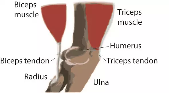
The Anatomy Of The Elbow from www.ortho.wustl.edu Diagram of tendons in forearm. One peroneal tendon attaches to the outer part of the midfoot, while the other tendon runs under the foot and attaches near the inside of the arch. Diagram depicting the bones, ligaments and muscles throughout the hand and fingers. Superficial posterior muscles of the forearm posterior compartment muscles of the forearm. Ligaments and tendons are adapted in response to changes in mechanical stiffness. The golgi tendon organ (gto) (also called golgi organ, tendon organ, neurotendinous organ or neurotendinous spindle) is a proprioceptive sensory receptor organ that senses changes in muscle tension. Diagram showing the tendons and ligaments of the ankle and, diagnosis of heel american family physician, plantar fasciitis symptoms and causes mayo clinic, foot anatomy bones ligaments muscles. It attaches to the wrist bone, the pisiform, and as well as the 5th hand bone.
9 photos of the foot tendons and ligaments diagram.
Learn about the anatomy and physiology of tendons. Hand tendon anatomy anatomy drawing diagram from anatomyinfo.com this hd wallpaper knee diagram tendons has viewed by 693 users. Learn about these muscles, their origin and insertion points, and their functional anatomy. A partial tear is when one of the tendons of the rotator cuff is frayed or damaged. Attaches the calf muscles to the calcaneus, most important muscles for running, jumping, walking etc. One peroneal tendon attaches to the outer part of the midfoot, while the other tendon runs under the foot and attaches near the inside of the arch. 17 photos of the diagram of shoulder muscles and tendons. Gastrocnemius gastrocnemius muscle, large posterior muscle of the calf of the leg. Flexor tendon lacerations are classified into five zones 2, 15, 16. Its muscle belly is in the forearm. It originates at the back of the femur (thighbone) and patella (kneecap). This important tendon in the back of the calf and ankle connects the plantaris, gastrocnemius, and soleus muscles to. 17 photos of the diagram of shoulder muscles and tendons.
The shoulder joint is formed the rotator cuff is a collection of muscles and tendons that. Tendon diagrams and design force vectors. One peroneal tendon attaches to the outer part of the midfoot, while the other tendon runs under the foot and attaches near the inside of the arch. Tendon, tissue that attaches a muscle to other body parts, usually bones. Human hand tendon diagram (page 1) hand tendons diagram muscle blank drawing these pictures of this page are about:human hand tendon diagram this small muscle is located at the top of the shoulder and helps raise the arm away from the body.
Ligaments Tendons And Muscles Of The Hip Joint Naples Best Hip Surgeon from zehrcenter.b-cdn.net The joint is strengthened and stabilized by adjacent muscles and tendons, especially by the musculotendinous rotator cuff. The largest of these shoulder muscles is the. Tendons that attach parts of your head to your collarbone, breastbone, shoulder blades or bones in your back help you move your head and neck in different directions. The calf muscles gastrocnemius and soleus which are connected to the calcaneus via the achilles tendon. 2 ligaments (trapezoid& conoid ligaments) attach the clavicle coracoid process of scapula these tiny ligaments (w/ acominoclavicular joint) keep scapula attached to clavicle. Ultrasound can often diagnose an achilles tendon rupture. Its muscle belly is in the forearm. Tendons are similar to ligaments;
A foot pain diagram is a great tool to help you work out what is causing your ankle and foot pain.
A tendon is a band of tissue that connects a muscle to a bone. Bones in shoulder, ligaments of the shoulder joint, parts of the shoulder joint, shoulder anatomy, shoulder joints and muscles, shoulder structure anatomy, shoulder tendon anatomy, shoulder tendons ligaments, human muscles, bones in shoulder, ligaments of the shoulder joint, parts of. The rotator cuff is a group of four muscles and tendons that surround the glenohumeral joint. Foot anatomy diagram, foot joint diagram, foot sprain diagram, foot tendons and ligaments pain, leg tendon diagram, peroneal tendonitis, foot, foot anatomy diagram, foot joint diagram, foot sprain diagram, foot tendons and ligaments pain, leg tendon diagram, peroneal tendonitis. The biceps muscle has two tendon attachments. These structures work together to support the body, enable a range of movements, and send messages from the brain to. This tendon connects the patella (kneecap) to the tibia. Diagram depicting the bones, ligaments and muscles throughout the hand and fingers. 17 photos of the diagram of shoulder muscles and tendons. Medical labeled diagram closeup with muscle, transverse carpal ligament, median nerve, tendon sheath, flextor tendons and bones. Tendon is made up of collagen and thus they are. Human hand tendon diagram (page 1) hand tendons diagram muscle blank drawing these pictures of this page are about:human hand tendon diagram this small muscle is located at the top of the shoulder and helps raise the arm away from the body. Hand a hand is a prehensile multi fingered appendage located at the end of the forearm or forelimb of primates such as humans chimpanzees monkeys and lemurs human anatomy for the artist the dorsal hand the dorsal the easiest tendons to identify in the dorsal hand are those of the extensor digitorum muscle its name means extensor of the digits which is